Introduction: thyroxine does not work in every third patient
According to cohort studies, 20–30% of patients receiving L-thyroxine replacement therapy at a biochemically adequate dose (TSH in the reference range) continue to have residual symptoms of hypothyroidism: fatigue, dry skin, weight gain, hair loss, brain fog, cold intolerance, tendency toward constipation, and reduced libido.
The standard cardiologist-endocrinologist response is: “TSH is normal; go home, the problem is not the thyroid.” But the problem is precisely in the thyroid axis: more specifically, thyroxine does not reach the cell in its active form.
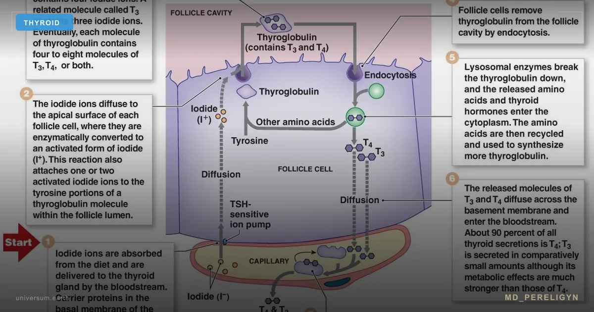
T4 (thyroxine) is a prohormone. By itself, it has almost no activity at the thyroid hormone receptor. The active form is T3 (triiodothyronine), whose affinity for the nuclear TR receptor is 10–15 times higher than that of T4. T4 → T3 conversion occurs in the periphery, not in the thyroid.
Key idea of the md_pereligyn protocol: normal TSH + normal fT4 + symptoms of hypothyroidism + low fT3 = functional hypothyroidism. This is not rare and not “patient laziness.” It is impaired peripheral conversion, visible in laboratory testing but not accounted for by the standard algorithm.
🌀
Physiology of T4 → T3 conversion
The conversion of T4 to T3 is catalyzed by three deiodinases, each with distinct biology:
▸D1 (type 1) — liver, kidneys. Produces most circulating T3; a selenium-dependent enzyme. ▸D2 (type 2) — brain, pituitary, muscle, brown adipose tissue. Local conversion for tissue T3; selenium-dependent. ▸D3 (type 3) — placenta, skin, fetal brain. Inactivates T4 into rT3 (reverse T3) and T3 into T2 — a “dead-end branch.”
All three deiodinases are selenoproteins. The enzyme active center contains selenocysteine. Without adequate selenium, the enzyme does not assemble or assembles incompletely.
Normal distribution: the thyroid secretes 80% T4 and 20% T3. Of this T4, peripheral conversion normally produces T3 — 30% (the physiological channel), and rT3 — 40% (physiological inactivation). The rest undergoes excretion and sulfation.
During stress, inflammation, or fasting, D2 switches toward D3 — conversion is directed into rT3. This is an evolutionary adaptation: during crisis, the body lowers metabolism. But with chronic stress, this switch can remain stuck in “winter” mode for years.
🌀
Where conversion fails: four mechanisms
•Selenium deficiency — deiodinase is a selenoprotein. Serum selenium levels <90 mcg/L are associated with reduced D1 and D2 activity. In regions with selenium-poor soil (Northern Europe, Northern Russia, Ukraine, Poland), deficiency is a background state. Therapeutic target: 120–150 mcg/L. •Chronically elevated cortisol — glucocorticoids directly activate D3 and suppress D2 at the transcriptional level. Result: T4 is diverted into the rT3 dead end, tissue T3 falls, and TSH remains normal (because fT4 does not change). Triggers: chronic stress, sleep deprivation <6 h, irregular eating, overtraining. •Ferritin deficiency — TPO (thyroid peroxidase) and conversion itself depend on iron as a cofactor. With ferritin <70 ng/mL, conversion suffers even with normal hemoglobin. Target ferritin in women with hypothyroidism: 70–100 ng/mL, not the “lower limit of normal.” •Systemic inflammation — IL-6, TNF-α, and IL-1β induce D3 and suppress D2. Marker: hsCRP >3 mg/L. Sources: visceral obesity, IBS, leaky gut, chronic infection (EBV, H. pylori), periodontitis, undiagnosed autoimmune disease.
Often all four factors operate in parallel in the same patient. Treating with selenium alone while cortisol remains elevated and ferritin remains low gives only a partial effect — hence “I tried selenium, it did not help.”
🌀
Markers: what to measure besides TSH
Standard screening (TSH + fT4) is blind to functional hypothyroidism. Expanded panel:
▸TSH (thyroid-stimulating hormone) — target 1.0–2.0 mIU/L on replacement therapy. The laboratory range 0.4–4.0 is too broad for symptomatic patients. ▸fT4 (free thyroxine) — target upper half of the reference interval (for example, 14–18 pmol/L with a reference range of 9–22). ▸fT3 (free triiodothyronine) — the main marker of tissue activity. Target range 4.0–7.0 pmol/L (or 3.5–6.5 with a reference range of 3.1–6.8). In functional hypothyroidism, fT3 is “pressed toward the lower limit.” ▸rT3 (reverse T3) — indicator of “dead-end” conversion. Target 9–24 ng/dL (or 0.14–0.54 nmol/L). >24 ng/dL suggests chronic stress or systemic inflammation. ▸fT3 / rT3 ratio — functional index. <0.2 (in nmol/nmol) indicates impaired conversion. >0.4 is normal. ▸Anti-TPO (thyroid peroxidase antibodies) — normal <35 IU/mL. Any elevation indicates a Hashimoto background, a common underlying contributor to symptoms. ▸Serum selenium — target 120–150 mcg/L. ▸Ferritin — target 70–100 ng/mL in women with hypothyroidism, 100–200 in men. ▸Zinc, serum iron, TIBC, transferrin saturation — expanded iron status. ▸Vitamin D 25(OH)D — target 60–80 ng/mL. ▸hsCRP — inflammation marker, target <1 mg/L. ▸4-point salivary cortisol (08:00, 12:00, 16:00, before sleep) — circadian rhythm; a single morning sample misses chronic stress.
🌀
Holistic protocol for restoring conversion
Principle: repair the enzyme, do not increase the T4 dose. Simply escalating L-thyroxine while the conversion block persists raises rT3 even further — the paradox of “more T4 = less T3.”
### 1. Selenium — the foundation
▸Selenium (L-selenomethionine) 200 mcg/day in the morning on an empty stomach, for at least 8 weeks, then daily or 5 days per week. ▸Check serum level after 8–12 weeks, target 120–150 mcg/L. ▸Do not exceed 400 mcg/day — toxicity (hair loss, brittle nails, garlic odor). ▸In Northern Europe and regions with selenium-poor soil, lifelong nutraceutical supplementation is justified.
### 2. Iron to target ferritin
▸Iron bisglycinate 25 mg every other day with vitamin C 500 mg for absorption. ▸No dairy, tea, or coffee within ±1 hour of intake. ▸Target ferritin 70–100 ng/mL; monitor every 8 weeks. ▸If oral forms are not tolerated, intravenous iron in an inpatient setting.
### 3. Zinc, magnesium, vitamin D
▸Zinc (bisglycinate / picolinate) 15–25 mg in the evening — cofactor for T3 synthesis and D1 regulation. ▸Magnesium (glycinate / taurate) 300–400 mg in the evening — thyroid receptor cofactor. ▸Vitamin D3 4000–10000 IU to a level of 60–80 ng/mL, + K2 (MK-7) 100–200 mcg. ▸Iodine is a separate topic; high doses are contraindicated in an autoimmune thyroiditis background. See the article Iodine and thyroid: 5 steps.
### 4. Lowering cortisol
▸Sleep 7–9 hours, bedtime before 23:00. One hour of sleep loss shifts cortisol by 15–20%. ▸Morning light exposure 10 minutes during the first hour after waking — circadian rhythm reset. ▸Adaptogens (ashwagandha 600 mg) — cortisol normalization and rT3 reduction in small studies. ▸Breathing practices 10 minutes/day — parasympathetic activation. ▸Limit caffeine after 14:00.
### 5. Reducing systemic inflammation
▸Mediterranean / DASH dietary pattern — olive oil, fish, vegetables, legumes. ▸EPA + DHA 2 g/day — omega-3, IL-6 reduction. ▸Curcumin (liposomal) 500 mg 2×/day — anti-inflammatory. ▸Treatment of hidden infections when confirmed (H. pylori, EBV reactivation, dental caries, periodontitis). ▸Reduction of visceral fat — waist circumference <94 cm (men) / <80 cm (women).
### 6. When to add T3 directly
▸Liothyronine (L-T3) 5–10 mcg in the morning or split dosing (5 in the morning + 5 at lunch) — discussed with a physician when conversion deficiency is confirmed and symptoms persist for 3–6 months on the full protocol. ▸NDT (natural desiccated thyroid) — a T4 + T3 combination in a physiological 4:1 ratio. Details are in the article Hypothyroidism and natural desiccated thyroid (NDT). ▸Check fT3, fT4, TSH after 6–8 weeks after starting T3-containing forms.
🌀
What does NOT work (and why)
▸Blindly increasing the L-thyroxine dose when fT3 is low — more substrate for D3, more rT3, same symptoms. ▸Selenium monotherapy with ferritin 15 ng/mL — the enzyme will not assemble without iron; the effect is partial. ▸Iodine at >500 mcg/day with selenium deficiency — provokes autoimmune thyroiditis exacerbation through oxidative stress in the thyrocyte. ▸T3 without cortisol correction — risks worsening anxiety and provoking tachycardia. ▸Self-prescribing liothyronine without an expanded panel and monitoring — uncontrolled T3 increases the risk of arrhythmias and osteoporosis. ▸“Normal TSH = thyroid is fine” — in a symptomatic patient with low fT3, this is a working hypothesis of functional hypothyroidism, not a reason to close the case. ▸Vegan / low-protein diet without correction — zinc, iron, B12, and tyrosine deficiency (T4 substrate). Conversion suffers.
🌀
When to seek care
▸TSH within reference range, fT4 within reference range, but fT3 <4.0 pmol/L or in the lower quartile of the reference range. ▸fT3 / rT3 ratio <0.2 (in nmol/nmol). ▸rT3 >24 ng/dL with normal TSH. ▸Residual hypothyroid symptoms on an adequate L-thyroxine dose for at least 6 months. ▸Suspected functional hypothyroidism without a formal diagnosis — symptomatic subclinical presentation. ▸Hashimoto background + progression of symptoms despite standard therapy. ▸Chronic stress, sleep disturbance, weight gain with a “normal” thyroid panel.
I run an expanded panel (TSH, fT4, fT3, rT3, anti-TPO, anti-Tg, selenium, ferritin, zinc, vitamin D, 4-point salivary cortisol, hsCRP) and create a personalized protocol for restoring conversion — without blindly increasing L-T4.
🌀
Conclusion
T4 is a prohormone. The active hormone is T3. The conversion enzyme — deiodinase — is assembled from selenium, iron, and zinc, and works under conditions of normal cortisol and low inflammation.
When any of these four conditions is disrupted, the patient enters functional hypothyroidism: TSH is normal, fT4 is normal, fT3 is low, and symptoms persist. This is visible on the expanded panel and invisible on the standard panel.
Treatment is directed not at the L-T4 dose, but at restoring the conditions required for the enzyme to work: selenium, iron, zinc, vitamin D, cortisol reduction, inflammation reduction, and, when needed, adding T3 or NDT. One measured fT3 can spare the dose escalation.
🌀
Sources
▸Bianco AC, et al. Paradigms of dynamic control of thyroid hormone signaling. *Endocr Rev* 2019;40:1000–1047. PMID 31033998 ▸Köhrle J. Selenium and thyroid. *Best Pract Res Clin Endocrinol Metab* 2009;23:815–827. PMID 19942156 ▸Wajner SM, Maia AL. New insights toward the acute non-thyroidal illness syndrome. *Front Endocrinol* 2012;3:8. PMID 22654852 ▸Salvatore D, et al. Thyroid hormones and skeletal muscle — new insights and potential implications. *Nat Rev Endocrinol* 2014;10:206–214. PMID 24322650 ▸Wiersinga WM. T4 + T3 combination therapy: an unsolved problem of increasing magnitude and complexity. *Endocrinol Metab* 2019;34:1–4. PMID 30912330 ▸Toulis KA, et al. Selenium supplementation in autoimmune thyroiditis. *Thyroid* 2010;20:1163–1173. PMID 20025778 ▸Mancini A, et al. Thyroid hormones, oxidative stress, and inflammation. *Mediators Inflamm* 2016;6757154. PMID 27051079
Related articles: Iodine and thyroid: 5 steps, Hypothyroidism and natural desiccated thyroid (NDT).
🌀
FAQ
Can thyroid function be assessed by TSH alone? No, not if symptoms are present. TSH is a marker of pituitary feedback, not tissue activity. Patients with residual symptoms on L-T4 need an expanded panel: fT4, fT3, rT3, anti-TPO, anti-Tg, selenium, ferritin, vitamin D, salivary cortisol.
What should be done if fT3 is low while TSH and fT4 are normal? This is a working hypothesis of functional hypothyroidism. Algorithm: check selenium, ferritin, zinc, vitamin D, cortisol, hsCRP. Correct identified deficiencies. After 8–12 weeks, repeat the expanded panel. If fT3 has not increased and symptoms remain, discuss T3 or NDT with a physician.
How quickly does selenium raise fT3? Clinically — 6–12 weeks at a dose of 200 mcg/day L-selenomethionine when baseline deficiency is present. Serum selenium rises within 8 weeks; fT3 rises more slowly because the enzyme must be synthesized, incorporated into the membrane, and build the T3 pool in tissues. Check at week 12.
Is rT3 dangerous by itself? rT3 is biologically inactive, but high rT3 is a marker of “winter” mode: chronic stress, inflammation, fasting, severe illness. rT3 falls in parallel with cortisol and hsCRP reduction — it is not treated in isolation.
When should T3 (liothyronine) or NDT be tried? After 3–6 months on the full protocol (selenium, iron, vitamin D, zinc, cortisol reduction) if low fT3 and symptoms persist. Starting low-dose T3 (5 mcg in the morning) or switching to NDT should be done only under physician supervision, with expanded panel monitoring after 6–8 weeks.
*This article is for informational purposes and does not replace medical consultation. Before starting any nutraceuticals, changing medication therapy, or undergoing diagnostic procedures, discuss the plan with your treating physician.*
🌀
The DIO2 Thr92Ala polymorphism: a genetic reason monotherapy fails
The original protocol lists four acquired mechanisms of conversion failure (selenium, cortisol, iron, inflammation) but does not address the most studied inherited mechanism. The type 2 deiodinase gene (DIO2) carries a common single-nucleotide polymorphism, rs225014, that substitutes alanine for threonine at codon 92 (Thr92Ala). Carrier frequency in unselected populations reaches 12–36% homozygous (Ala/Ala) and approximately 40–50% heterozygous, depending on ancestry PMID: 28324063.
The substitution does not abolish enzyme function — DIO2 still converts T4 to T3 — but it alters intracellular trafficking. Ala/Ala variant enzyme accumulates in the Golgi apparatus rather than the endoplasmic reticulum, where its physiological substrate is delivered. The result is reduced T3 generation at the tissue level despite normal serum T4 and normal-to-high TSH. Brain tissue, which depends almost entirely on local DIO2 activity for its T3 supply, is the most affected compartment PMID: 28855267.
Clinically, this matters because the standard "TSH-normalized" levothyroxine dose may produce adequate plasma T4 yet leave central nervous system T3 inadequate. The Weetman group's pharmacogenetic analysis of two combined-therapy trials found that Ala/Ala carriers had measurably better psychological well-being on combined L-T4 + L-T3 than on L-T4 monotherapy, whereas Thr/Thr carriers showed no preference PMID: 28324063. Subsequent meta-analyses have been mixed but consistently identify the Ala/Ala subgroup as the most plausible responder phenotype PMID: 35629879.
Practical implications for the patient who continues to feel hypothyroid on adequate L-T4:
- Genotyping (DIO2 rs225014) is available in most clinical genomics panels and is one-time, inexpensive, and does not require fasting. - The polymorphism does not predict who needs T3; it identifies a population in whom a 3–6 month trial of L-T4 + L-T3 (or NDT) is more likely to produce subjective improvement. - Ala/Ala status should not override fT3 measurement. If fT3 is mid-range or higher and rT3 is suppressed, peripheral conversion is intact regardless of genotype. - Genotype does not change TSH targets, monitoring frequency, or contraindications to T3.
The DIO2 polymorphism explains why two patients with identical TSH, fT4, ferritin and selenium can have divergent symptomatic responses to the same regimen. It is one mechanism — not a substitute for the metabolic workup outlined earlier — but it deserves a place in the diagnostic ladder when fT3 is low and the four acquired causes have been corrected.
🌀
Drugs that inhibit T4→T3 conversion: a pharmacological checklist
The article identifies cortisol as a conversion inhibitor but does not list the medications that produce the same biochemical effect. Drug-induced impairment is common, reversible, and often missed because the patient remains on chronic therapy.
Amiodarone. Iodine content (37% by weight) and direct DIO1/DIO2 inhibition lower fT3 and raise rT3 within 1–3 months of initiation. Approximately 14–18% of patients develop biochemical hypothyroidism; a smaller subset develops Type 1 or Type 2 amiodarone-induced thyrotoxicosis. fT3 may fall below the reference range while TSH remains normal for weeks. Monitoring: TSH, fT4, fT3 at baseline, 3 months, then every 6 months PMID: 2744218.
Glucocorticoids. Prednisolone ≥20 mg/day, dexamethasone ≥1 mg, and equivalent doses of hydrocortisone suppress DIO1 and stimulate DIO3, lowering fT3 and TSH and raising rT3. The effect appears within 48 hours and persists for the duration of therapy. Inhaled and topical steroids at standard doses do not produce systemic conversion changes. Endogenous Cushing syndrome reproduces the same pattern.
Propranolol (and to a lesser extent other non-selective beta-blockers). Doses ≥160 mg/day inhibit DIO1, reducing T3 generation by approximately 30%. The effect is dose-dependent and reverses within days of discontinuation. Cardioselective beta-blockers (atenolol, metoprolol, bisoprolol) at usual doses have minimal effect on conversion.
Propylthiouracil (PTU). Inhibits both thyroid peroxidase and DIO1. In Graves disease this is therapeutic; in a patient with conversion-pattern hypothyroidism it should not be used.
Iopanoic acid, ipodate, and iodinated radiographic contrast. Acutely block DIO1 and DIO2; effect on fT3 lasts 2–4 weeks after a single CT angiography or coronary angiogram. Lab interpretation should be deferred during this window.
Lithium. Inhibits thyroid hormone release and modestly suppresses peripheral conversion. Combined effect produces overt or subclinical hypothyroidism in 15–25% of long-term users.
Less recognized. High-dose salicylates and furosemide displace T4 from binding proteins and can transiently alter free hormone levels; clinical conversion impairment is minor at standard doses.
Before diagnosing conversion failure as the primary problem, the medication list must be reviewed. Discontinuing or switching a confounding drug — when safe — is faster and more specific than any micronutrient protocol.
🌀
Liothyronine pharmacokinetics and a stepwise titration protocol
The article suggests "5–10 mcg" of liothyronine without specifying half-life, peak timing, or titration. These details determine whether T3 therapy succeeds or produces palpitations and tremor.
Oral liothyronine is rapidly and almost completely absorbed (bioavailability ~95%). Peak serum T3 occurs 2–4 hours after ingestion, with the post-dose Cmax frequently exceeding the upper reference limit even at therapeutic doses. The elimination half-life is approximately 20–24 hours, but the biological half-life — the duration of receptor occupancy — is shorter, which is why a single morning dose produces visible serum oscillation and symptomatic swings PMID: 23072197.
Practical consequences:
- A single morning 10 mcg dose produces a supraphysiologic peak at 09:00–11:00 and a trough by the next morning. Split dosing (2.5–5 mcg twice daily) attenuates the peak and provides smoother coverage. - Trough fT3 (drawn before the next dose) is the only meaningful monitoring value. Peak fT3 (drawn 2–4 hours after dosing) is uninterpretable because it captures the absorption surge, not the steady state. - Starting dose in a patient already on adequate L-T4 with low fT3: 2.5–5 mcg in the morning, with a parallel reduction of L-T4 by 12.5–25 mcg to keep TSH stable. The conversion-equivalence is roughly 1 mcg T3 ≈ 3 mcg T4. - Titrate at 4–6 week intervals using TSH, fT4, and pre-dose fT3. Target: TSH within reference, fT3 in the upper third of reference, fT4 mid-reference or slightly below.
Stopping rules. Discontinue or reduce T3 if any of the following occur: resting heart rate >100 bpm sustained for 48 hours, new atrial fibrillation, palpitations with documented ectopy, weight loss exceeding 2 kg/month without intent, suppressed TSH below 0.1 mIU/L on repeat testing, or new bone density decline in postmenopausal women.
Contraindications and cautions. Untreated adrenal insufficiency (precipitates adrenal crisis), recent acute coronary syndrome (<6 weeks), known atrial fibrillation without rate control, severe osteoporosis without bisphosphonate cover, and pregnancy (use L-T4 only — T3 does not cross the placenta efficiently and fetal brain development depends on maternal T4 → fetal T3 conversion).
The largest combination-therapy trials and a 2024 systematic review found no consistent group-level benefit of L-T4 + L-T3 over L-T4 alone but identified subgroups with subjective improvement, particularly patients with persistent symptoms despite normalized TSH PMID: 39290156, PMID: 38124252. T3 therapy is therefore an n-of-1 trial with defined start, stop, and monitoring criteria, not a long-term default.
🌀
Why low T3 matters: outcomes data beyond symptoms
The article frames low fT3 as a cause of fatigue and metabolic slowdown but does not cite the outcomes literature. Low fT3 is a measured prognostic marker in several non-thyroid conditions.
In chronic heart failure, fT3 below the lower reference limit is an independent predictor of all-cause and cardiovascular mortality after adjustment for ejection fraction, NT-proBNP, and NYHA class. The Pisa cohort and subsequent meta-analyses report hazard ratios of 1.5–2.4 for cardiovascular death in low-T3 patients with stable heart failure PMID: 37096279, PMID: 31806208.
In acute myocardial infarction, low admission T3 correlates with infarct size, lower left ventricular ejection fraction at discharge, and higher 1-year mortality. The mechanism is presumed to be reduced cardiomyocyte T3 signaling at a time of maximal repair demand.
In critical illness, low T3 with normal or low TSH is the hallmark of non-thyroidal illness syndrome. Mortality in ICU populations rises with the magnitude of fT3 suppression. This is associated, not causal — replacing T3 in unselected critically ill patients has not improved survival in randomized trials, and current consensus is observation, not treatment PMID: 33320308.
In type 2 diabetes and metabolic syndrome, lower fT3/fT4 ratios correlate with higher HOMA-IR, higher visceral adiposity, and higher fasting glucose. The relationship is bidirectional: insulin resistance reduces DIO2 expression in skeletal muscle and adipose tissue, and lower local T3 reduces glucose uptake and mitochondrial biogenesis PMID: 38201918.
Two points for the ambulatory patient:
- A low-normal fT3 in a euthyroid patient with cardiovascular disease, diabetes, or unexplained fatigue is not a benign finding. It does not automatically mandate T3 replacement, but it does identify a population worth investigating for the four acquired conversion causes already discussed. - The outcome data refer to spontaneous low T3, not iatrogenic low T3 from levothyroxine monotherapy. Whether correcting iatrogenic low T3 with combined therapy improves cardiovascular outcomes is an open question; trials have measured symptoms and weight, not mortality.
🌀
Differential diagnosis: impaired conversion versus three look-alikes
The phrase "functional hypothyroidism" risks being applied to any patient with a low fT3, including patients whose physiology is entirely different. Four entities share the biochemical pattern of low or low-normal fT3, and each has a different management.
1. Impaired peripheral conversion (the subject of this article). TSH normal, fT4 normal or high-normal, fT3 low, rT3 high, fT3/rT3 ratio reduced. Ferritin, selenium, zinc, hsCRP, cortisol provide the mechanistic explanation. Treatment: correct deficiencies; consider T3 trial if symptoms persist.
2. Non-thyroidal illness syndrome (sick euthyroid). Acute or chronic systemic illness, often with measurable inflammation (hsCRP elevated, ferritin sometimes elevated as an acute-phase reactant). TSH may be low, normal, or transiently elevated during recovery. fT4 typically low-normal, fT3 low, rT3 high. Caloric restriction reproduces a similar pattern as an adaptive response — fT3 falls and rT3 rises within 48–72 hours of fasting and reverses within days of refeeding PMID: 25349245. Treatment: the underlying illness, not thyroid hormone PMID: 33320308.
3. Central (secondary) hypothyroidism. Pituitary or hypothalamic disease. TSH is low or inappropriately normal for the low fT4. fT3 follows fT4 downward. Distinguished from impaired conversion by the low fT4. Imaging (pituitary MRI), other anterior pituitary axes (cortisol, ACTH, IGF-1, gonadotrophins, prolactin) must be evaluated before any thyroid hormone is started. Levothyroxine is dosed to mid-reference fT4, not by TSH.
4. Thyroid hormone resistance (RTH). THRB or THRA mutation. TSH normal or high, fT4 and fT3 both elevated, symptoms variable. Often misdiagnosed as Graves disease. Family history typically positive. Genetic testing confirms. No T3 indicated.
A pragmatic decision rule:
- Low fT3 + low fT4 + low or low-normal TSH → think central hypothyroidism, request pituitary workup. - Low fT3 + normal fT4 + normal TSH + acute illness → think non-thyroidal illness, treat the illness. - Low fT3 + normal fT4 + normal TSH + chronic symptoms + high rT3 + nutrient/cortisol abnormality → impaired conversion. - High fT3 and fT4 with non-suppressed TSH → think resistance, refer to endocrinology.
Selenium supplementation has been shown to reduce TPO-antibody titers in Hashimoto thyroiditis in several randomized trials PMID: 38243784, PMID: 33650299, but it does not treat any of the four entities above by itself. It corrects one of several conversion cofactors; the rest of the differential still applies.
The diagnostic separation is not academic. The patient with central hypothyroidism started on T3 without cortisol assessment can precipitate an adrenal crisis. The patient with non-thyroidal illness given thyroid hormone can develop arrhythmia without any benefit. The patient with resistance treated as conversion failure receives medication that worsens the resistance state. Functional hypothyroidism is a real entity but a residual diagnosis — made after the alternatives have been excluded, not before.
References
- Bianco AC, et al. Paradigms of dynamic control of thyroid hormone signaling. Endocr Rev 2019;40:1000–1047. PMID 31033998
- Köhrle J. Selenium and thyroid. Best Pract Res Clin Endocrinol Metab 2009;23:815–827. PMID 19942156
- Wajner SM, Maia AL. New insights toward the acute non-thyroidal illness syndrome. Front Endocrinol 2012;3:8. PMID 22654852
- Salvatore D, et al. Thyroid hormones and skeletal muscle — new insights and potential implications. Nat Rev Endocrinol 2014;10:206–214. PMID 24322650
- Wiersinga WM. T4 + T3 combination therapy: an unsolved problem of increasing magnitude and complexity. Endocrinol Metab 2019;34:1–4. PMID 30912330
- Toulis KA, et al. Selenium supplementation in autoimmune thyroiditis. Thyroid 2010;20:1163–1173. PMID 20025778
- Mancini A, et al. Thyroid hormones, oxidative stress, and inflammation. Mediators Inflamm 2016;6757154. PMID 27051079
- PMID: 28324063. PMID 28324063
- PMID: 28855267. PMID 28855267
- PMID: 35629879. PMID 35629879
- PMID: 2744218. PMID 2744218
- PMID: 23072197. PMID 23072197
- PMID: 39290156. PMID 39290156
- PMID: 38124252. PMID 38124252
- PMID: 37096279. PMID 37096279
- PMID: 31806208. PMID 31806208
- PMID: 33320308. PMID 33320308
- PMID: 38201918. PMID 38201918
- PMID: 25349245. PMID 25349245
- PMID: 38243784. PMID 38243784
- PMID: 33650299. PMID 33650299