Introduction: Starvation Is Not Weight Loss
When you aggressively cut calories, fast, and live in stress, the body does not turn on fat burning — it turns on survival mode. This is not "weak willpower" or "bad genetics" — this is a normal physiological response to threat.
First water leaves and it seems to work. Then reverse T3 (rT3) rises — an inhibitory metabolite that slows energy expenditure. Active T3 falls, mitochondria slow, you feel cold, swollen, foggy. Weight stalls or creeps up.
Then comes the classic complaint: "I barely eat — why am I not losing weight?" Because starvation is a signal to the body to conserve, not spend.
This article breaks down the mechanism of this trap and the five-step thyroid work to restore active metabolism.
🌀
Mechanism: Reverse T3 as the Brake Pedal
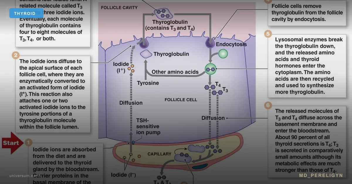
T4 (thyroxine) is a "prohormone" that tissues convert into active T3 or inactive reverse T3 (rT3). The T3/rT3 ratio is determined by deiodinase enzymes:
▸Deiodinase 1 and 2 (D1, D2) — convert T4 to T3 (active) ▸Deiodinase 3 (D3) — converts T4 to rT3 (brake)
Normally the balance is shifted toward T3. Under stress — fasting, illness, inflammation, surgery, sleep deprivation — balance shifts sharply toward rT3. This is evolutionary: the body conserves energy to survive famine.
The problem: in the modern world, chronic caloric deficit, chronic stress, and chronic inflammation keep people in high-rT3 mode for years. This is the syndrome of "untreated functional hypothyroidism with normal labs".
Labs show:
▸TSH — normal or slightly elevated ▸T4 — normal ▸fT3 — low-normal or below range ▸rT3 — elevated ▸fT3/rT3 ratio < 0.2 (normal > 0.2)
A standard endocrinologist looks at TSH and T4, says "all good". But clinically the patient has hypothyroidism: fatigue, cold intolerance, edema, hair loss, weight gain.
🌀
What Causes rT3 to Rise
▸Prolonged caloric deficit (more than 4–6 weeks at significant deficit) ▸Very low-carbohydrate diets without adjustment ▸Chronic stress — high cortisol suppresses D2 ▸Inflammation (elevated CRP, TNF-α) ▸Sleep deprivation less than 6 hours ▸Selenium deficiency — critical for D2 ▸Iron deficiency — ferritin < 70 ng/mL ▸Heavy training without recovery ▸Fatty liver disease — liver does 60% of conversion
Each factor raises rT3 by 20–40%. In combination the effect is multiplicative.
🌀
Five Stages of Thyroid Work for Weight Loss
This is the key idea of the md_pereligyn protocol: before "going on a diet", verify thyroid functionality at five levels. This works both ways: prevents loss of thyroid function during deficit, and helps recover if the trap has already triggered.
### 1. Iodine Cellular Uptake (NIS Symporter)
Full breakdown of all 5 iodine stages — in Iodine and Thyroid: 5-Step Protocol. Here — brief overview in weight-loss context.
Iodine must enter the thyroid cell via the sodium-iodide symporter. This requires:
▸Normal sodium-potassium gradient ▸Cellular energy ▸Living, non-inflamed tissue
What helps: avoid burnout, chronic stress, maintain cellular energy. Fasting breaks NIS — explaining why aggressive diets always lead to functional hypothyroidism.
Nutraceuticals: magnesium 300–400 mg, electrolytes, protein 1.2–1.5 g/kg.
### 2. Thyroid Peroxidase (TPO)
Without TPO, iodine will not oxidize and incorporate into hormones. Critical here are ferritin (target 70–100, not "normal range") and anti-TPO status.
Nutraceuticals: iron bisglycinate if ferritin < 70, vitamin C 500 mg for absorption. With elevated anti-TPO — selenium + vitamin D + anti-inflammatory diet.
### 3. Peroxide Protection
During thyroid hormone synthesis the gland generates hydrogen peroxide. Without selenium, peroxide damages tissue from within. This is critical in Hashimoto patients.
Nutraceuticals: selenium 100–200 µg/day, NAC 600–1200 mg, glycine 3 g, vitamin C.
### 4. Iodine + Iodide (Systemic Context)
Iodine is needed, but without selenium, iron, and proper environment, it can do harm. Blind iodine with cofactor deficiencies worsens Hashimoto. md_pereligyn principle:
1. Labs first (full panel + ferritin + selenium) 2. Cofactor preparation 4–8 weeks 3. Only then iodine, starting with microdoses 4. Recheck at 8–12 weeks
### 5. T4 → T3 Conversion
This is where it is decided whether you have energy or just "normal T4" in labs. Conversion is broken by:
▸Fasting (prolonged deficit) ▸Inflammation ▸Liver (fatty liver disease) ▸Gut (20% of conversion) ▸Selenium, zinc, iron deficiencies
What helps: stop fasting, sleep, protein, bile, gut, stress management.
Nutraceuticals: selenium 200 µg, zinc 15–25 mg, magnesium 300–400 mg, omega-3 1–2 g, tyrosine 500 mg morning.
🌀
How to Escape the rT3 Trap
If rT3 is already elevated and you are in survival mode, you cannot continue aggressive deficit — this deepens the problem. Recovery plan:
### Step 1: Reverse-diet 4–8 weeks
Gradually increase calories by 100–200 kcal/week up to maintenance. The body stops perceiving the situation as famine. Metabolism recovers. Paradox: to lose weight, you first have to start eating normally.
### Step 2: Cofactor Recovery
Selenium 200 µg + zinc 25 mg + magnesium 400 mg + iron if ferritin < 70 + vitamin D to 60–80 ng/mL + omega-3 EPA/DHA 1–2 g.
### Step 3: Sleep and Stress
7–9 hours of sleep. Cortisol elevated — that is a conversion blocker. Adaptogens (rhodiola, ashwagandha) can help, but primary focus is regimen.
### Step 4: Liver and Gut
60% of conversion in liver, 20% in gut. Bile support (ox bile, milk thistle), butyrate, probiotics, protein 1.2–1.5 g/kg.
### Step 5: Recheck and Continue
At 8–12 weeks recheck: TSH, fT4, fT3, rT3, fT3/rT3 ratio. If ratio > 0.2 — system is recovering, can enter moderate deficit for fat loss.
🌀
What DOES NOT Work (and Why)
▸Pure caloric deficit without cofactor support — deepens survival mode ▸T3 supplementation bypassing the system — short-term improvement followed by crash ▸"I just won't eat" — after 14 days of significant deficit, rT3 rises 30–50% ▸Cardio + deficit — powerfully amplifies cortisol and rT3 when applied together ▸Aggressive keto without adjustment — in some patients spikes rT3 in first 4 weeks
🌀
What DOES Work
▸Reverse-diet to escape survival mode ▸Resistance training (raises T3 via muscle mass), not cardio ▸Full endocrine diagnostics before any diet ▸Cofactor preparation 4–8 weeks before deficit ▸Moderate deficit 10–20% below maintenance, no more ▸Adequate protein (minimum 1.2 g/kg) ▸Sleep 7–9 h — sleep loss raises rT3 in 2 nights ▸Stress management — cortisol and rT3 are directly linked
🌀
Principle
"I barely eat — why am I not losing weight?" is not a psychology or motivation question. It is a physiology question.
The body does not distinguish between "diet for weight loss" and "famine during war". The response is the same: conserve energy, accumulate reserves, slow metabolism. And rT3 is the primary molecular instrument of this response.
Modern endocrinology (Bauer M. & Whybrow PC., *Thyroid*, 2014) recognizes rT3 as a functional antagonist of active T3 and proposes it as a marker of "untreated functional hypothyroidism". Most clinics do not run this panel — explaining why millions of women diet for years without results.
🌀
Conclusion
Before going on yet another diet — verify whether you are already in survival mode. Full thyroid panel (TSH + fT4 + fT3 + rT3 + anti-TPO) + ferritin + vitamin D + selenium gives the complete picture.
Weight loss is not a question of "eat less, move more". It is a question of maintaining metabolism in active state during moderate deficit. Without this foundation, any diet is a trap.
---
References:
- Bauer M, Whybrow PC. *Thyroid Hormones and Mood: A Reappraisal of the Reverse T3 Syndrome.* Thyroid 2014
- Rosenbaum M et al. *Long-term persistence of adaptive thyroid response to weight loss.* PMID 18996896
- Fontana L et al. *Long-term low-protein, low-calorie diet and endurance exercise modulate metabolic factors.* PMID 17389712
- Müller MJ et al. *Adaptive thermogenesis with weight loss in humans.* PMID 22968145
🌀
Laboratory reference ranges for reverse T3 and the fT3/rT3 ratio
The article specifies the fT3/rT3 ratio cutoff (>0.2) without unit definitions or analytical caveats. This section closes that gap.
Reverse T3 is most often reported in two units: ng/dL (US laboratories) and pmol/L (European laboratories). Adult reference intervals are method-dependent and vary across assays, but commonly cited ranges are:
- LC–MS/MS or immunoassay (US): 9.2–24.1 ng/dL - pmol/L conversion: divide ng/dL by 6.5; reference 14–37 pmol/L
For the fT3/rT3 ratio the calculation requires both values in pg/mL (free T3) and ng/dL (rT3) converted to a common scale, or both as molar concentrations (pmol/L). The most commonly used clinical formula is fT3 (pg/mL) × 100 / rT3 (ng/dL), with the published cutoff for adequate peripheral conversion at >0.20, optimal >0.25 PMID: 24847342. Ratios below 0.15 are consistent with significant impairment of T4-to-T3 conversion in the absence of overt thyroid disease PMID: 18510706.
Analytical caveats limit the diagnostic usefulness of an isolated rT3 measurement:
- rT3 has a half-life of approximately 5 hours and is extremely sensitive to acute stressors. A single elevated value within 48 hours of surgery, fasting longer than 24 hours, an acute infection, or strenuous exercise reflects transient deiodinase shift, not chronic dysfunction. - Hemolysis falsely elevates rT3 in older immunoassays. - Heparin (including low-molecular-weight heparin used for venous access) artifactually raises free T4 and may distort the calculated ratio. - Diurnal variation is small but real; standardize sampling between 08:00–10:00 fasting.
For diagnostic certainty the panel should be drawn twice, 4–6 weeks apart, both samples in the early morning fasting state, off all non-essential supplements for 72 hours. A single elevated rT3 in isolation does not justify intervention.
Free T3 reference ranges (2.3–4.2 pg/mL or 3.5–6.5 pmol/L in most assays) should be interpreted relative to the upper-mid quartile of the reference interval, not simply "in range" PMID: 18852350. A free T3 in the bottom 20% of the reference range with elevated rT3 is the laboratory phenotype of the trap described in the article.
🌀
Cortisol–deiodinase mechanism: how stress flips the conversion switch
The article states that "high cortisol suppresses D2" without describing the molecular pathway or providing measurable cortisol thresholds. This section addresses that gap.
Glucocorticoids exert dual inhibition on the peripheral thyroid hormone economy. First, cortisol directly suppresses transcription of the DIO2 gene encoding deiodinase 2, the principal enzyme generating active T3 from T4 in pituitary, brown adipose tissue, skeletal muscle, and brain. The glucocorticoid receptor binds a negative response element in the DIO2 promoter region, downregulating expression within 4–6 hours of sustained cortisol elevation PMID: 11836274. Second, glucocorticoids simultaneously upregulate DIO3, the enzyme converting T4 to rT3, in liver and skeletal muscle PMID: 16148345. The combined effect is a coordinated shift from active to inactive thyroid hormone within hours of physiological stress.
Cortisol also suppresses TRH release from the hypothalamus, lowering TSH output. This is why a stressed patient frequently presents with TSH at the lower end of the reference range despite clear clinical hypothyroidism — TSH is artifactually suppressed by central HPA-axis tone, not by adequate peripheral hormone PMID: 9024227.
Measurable thresholds clinicians can use:
- Morning serum cortisol >18 µg/dL (497 nmol/L) outside acute illness suggests sustained HPA activation - Diurnal salivary cortisol with elevated evening (22:00) value >0.2 µg/dL indicates loss of the normal nocturnal nadir - 24-hour urinary free cortisol >50 µg/24h confirms systemic glucocorticoid excess - Late-night salivary cortisol is the most sensitive screen for chronic stress-driven HPA dysregulation
Importantly, the relationship is non-linear. A short cortisol spike from acute exercise or a single stressful event does not produce sustained rT3 elevation. Chronic cortisol elevation across days to weeks is required to establish the high-rT3, low-fT3 phenotype PMID: 21115738. This explains the article's clinical observation that escape from the trap requires sleep and stress regulation before nutritional rehabilitation will normalize the conversion ratio.
🌀
Differential diagnosis: non-thyroidal illness syndrome versus diet-induced functional hypothyroidism
The article describes one mechanism — caloric-restriction-induced rT3 elevation — without distinguishing it from non-thyroidal illness syndrome (NTIS, also called euthyroid sick syndrome or low-T3 syndrome). The two share an identical laboratory signature but require opposite management.
NTIS is the systemic adaptive response to acute or critical illness: sepsis, myocardial infarction, major trauma, hospitalization in intensive care, advanced malignancy, decompensated heart failure, or chronic renal disease PMID: 15049952. The pattern is suppressed T3, normal or low T4, normal or low TSH, and elevated rT3. Free T3 may fall below 1.5 pg/mL in severely ill patients. The mechanism overlaps with caloric-restriction physiology — cytokine-driven D3 upregulation, suppressed D1 and D2 — but the trigger is illness, not diet.
Clinical distinction:
- NTIS: clear precipitating illness, often hospitalized, elevated inflammatory markers (CRP >10 mg/L, IL-6 elevated), prognostic marker rather than treatment target. Thyroid hormone replacement in NTIS does not improve outcomes and may worsen mortality in cardiac patients [PMID: 8636404](https://pubmed.ncbi.nlm.nih.gov/8636404/). - Diet-induced functional hypothyroidism: ambulatory patient, prolonged caloric deficit or restrictive diet history, normal or mildly elevated CRP, symptoms of fatigue and weight stall, no acute illness. The fT3/rT3 ratio responds to reverse-diet and cofactor repletion within 8–12 weeks.
Additional differentials that present with a similar phenotype:
- Subclinical hypothyroidism with autoimmune thyroiditis — distinguished by elevated anti-TPO and TSH >4.5 mIU/L - Pituitary insufficiency (central hypothyroidism) — low TSH with low T4, often with low cortisol and gonadotropins; requires MRI - Polymorphisms in DIO2 (Thr92Ala variant) — present in approximately 16% of populations, associated with persistent low-grade fT3 deficiency despite normal labs [PMID: 25540980](https://pubmed.ncbi.nlm.nih.gov/25540980/)
Before initiating the protocol described in the article, exclude NTIS by reviewing recent illness history, inflammatory markers, and clinical context. A patient with active infection, acute heart failure, or recent surgery should not undergo reverse-diet intervention until the underlying illness is resolved.
🌀
Drug-induced reverse T3 elevation
The article lists lifestyle drivers of rT3 elevation but omits iatrogenic causes. Several common medications shift the deiodinase balance and may explain treatment-resistant cases.
Beta-blockers, particularly propranolol at doses above 160 mg/day, inhibit D1 activity and impair peripheral T4-to-T3 conversion. Lower doses and cardioselective agents (metoprolol, bisoprolol) have less effect. Atenolol at standard antihypertensive doses produces minimal interference PMID: 32593258.
Amiodarone is the most potent pharmacologic cause of rT3 elevation. The drug contains 37% iodine by weight, has a half-life of 50–100 days, inhibits both D1 and D2, and blocks T3 binding to nuclear receptors. Patients on amiodarone routinely show rT3 two to three times the upper reference limit; this is expected and not pathologic unless accompanied by clinical symptoms. Amiodarone-induced thyroid dysfunction (both hyper- and hypothyroid forms) affects 15–20% of patients on long-term therapy and requires specialist management PMID: 24432020.
Glucocorticoids in any form — oral prednisone above 10 mg/day, inhaled corticosteroids at high doses, intra-articular injections, and topical clobetasol on large surface areas — produce the same deiodinase shift as endogenous cortisol described above. Patients on chronic glucocorticoid therapy for autoimmune disease, asthma, or inflammatory bowel disease frequently present with the rT3 phenotype.
Iodinated radiographic contrast media (used in CT angiography, coronary angiography) deliver a large iodine bolus and transiently inhibit D1 for 4–8 weeks. Thyroid panels drawn within 6 weeks of contrast exposure are unreliable.
Other relevant agents:
- Phenytoin and carbamazepine — induce hepatic clearance of T4, lower total T4, secondarily reduce conversion - Lithium — inhibits thyroid hormone release; chronic users develop hypothyroidism in 20–30% of cases - SSRIs (sertraline, fluoxetine) — modest TSH elevation in some patients, mechanism unclear - Oral estrogens — increase thyroxine-binding globulin, raise total T4 without changing free fractions; this is a binding artifact, not a true conversion problem
Before applying the reverse-diet protocol, document the patient's full medication list and consider whether dose adjustment, drug holiday, or substitution of an alternative agent is feasible in consultation with the prescribing physician.
🌀
Resistance training protocol for thyroid recovery
The article recommends resistance training over cardio for rT3 reduction without specifying volume, intensity, or frequency. This section provides the evidence-based parameters.
Resistance training raises peripheral T3 via three mechanisms: increased skeletal muscle mass expands the largest D2-expressing tissue compartment; mechanical loading upregulates DIO2 expression in trained muscle; and exercise-induced lactate increases hepatic D1 activity. The net effect is a higher fT3 and lower fT3/rT3 ratio impairment at any given caloric intake.
Protocol parameters supported by randomized data:
- Frequency: 3 sessions per week, non-consecutive days. Higher frequency (5–6 sessions) is counterproductive in the rT3-elevated patient because it increases cortisol exposure without proportional anabolic benefit. - Volume: 10–15 working sets per major muscle group per week, distributed across sessions. Total session duration 45–60 minutes including warm-up. - Intensity: compound movements at 70–85% of one-repetition maximum, 6–10 repetitions per set, 2–3 minutes rest between sets. This intensity range maximizes growth hormone and IGF-1 response, which independently support T3 conversion. - Exercise selection: squat or leg press, deadlift or hip hinge, horizontal press (bench press or dumbbell press), vertical pull (pull-up or pulldown), horizontal pull (row), and one or two accessory movements per session. - Progression: linear progression in load by 2.5–5% per week as tolerated, with deload week every 4–6 weeks (50% volume) to limit cortisol accumulation.
Cardiovascular work is not contraindicated but should be capped at 60–90 minutes per week of zone-2 intensity (60–70% maximum heart rate). Higher-intensity interval training and steady-state cardio above 70% maximum heart rate exceeding 2 hours per week elevates cortisol enough to attenuate the resistance-training benefit on T3 conversion.
Sleep timing matters as much as training timing. Training within 4 hours of intended bedtime delays sleep onset in many patients and blunts the nocturnal cortisol nadir. Morning or early-afternoon sessions are preferred. Post-workout protein intake of 0.3 g/kg within 90 minutes supports muscle protein synthesis without producing the cortisol response seen with prolonged post-exercise fasting.
Expected timeline: measurable lean mass gain of 0.5–1 kg per month in deficit-controlled patients, with fT3/rT3 ratio improvement detectable at 8–12 weeks. The protocol is not optional adjunct — in patients refractory to nutritional repletion alone, structured resistance training is frequently the variable that finally normalizes the conversion ratio.
References
- PMID 18996896. PMID 18996896
- PMID 17389712. PMID 17389712
- PMID 22968145. PMID 22968145
- PMID: 24847342. PMID 24847342
- PMID: 18510706. PMID 18510706
- PMID: 18852350. PMID 18852350
- PMID: 11836274. PMID 11836274
- PMID: 16148345. PMID 16148345
- PMID: 9024227. PMID 9024227
- PMID: 21115738. PMID 21115738
- PMID: 15049952. PMID 15049952
- PMID: 8636404. PMID 8636404
- PMID: 25540980. PMID 25540980
- PMID: 32593258. PMID 32593258
- PMID: 24432020. PMID 24432020